


What are the advantages of a spectral autofocus microscopy system?
The spectral autofocus microscopy system offers high-precision autofocus capabilities, effectively minimising human error and significantly improving experimental efficiency. It is also capable of adapting to a wide range of complex sample imaging scenarios, making it a key piece of equipment for the intelligent upgrading of microscopy experiments.

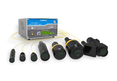
In traditional microscopy experiments, focusing has always been a key bottleneck affecting image quality. Conventional manual focusing relies heavily on the operator’s experience and tactile sensitivity. Not only does it require repeated fine-tuning of the lens focus—a highly time-consuming process—but it is also highly susceptible to issues such as focus drift and uneven sharpness caused by human visual bias and inconsistent application of force. This results in blurred images and invalid data; particularly in long-duration, high-throughput experiments, human error accumulates over time, severely compromising the accuracy of experimental results.

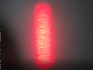
The spectral autofocus microscopy system, however, has completely overcome the limitations of traditional equipment. Based on the core principle of spectral detection, the system captures the focal length signals of the sample image in real time, automatically completing the entire process—from focus recognition and parameter fine-tuning to precise positioning—without the need for manual intervention. Unlike the subjectivity of manual focusing, the system’s focusing logic is consistent and its response stable, thereby eliminating at source any errors caused by human operation and ensuring uniform standards for every image.

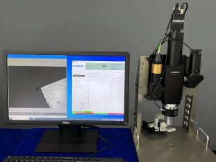
At the same time, its powerful adaptability perfectly resolves the challenges of imaging complex samples. When faced with special samples that are uneven, multi-layered or have uneven light transmittance, traditional microscopy equipment is highly prone to localised defocusing and imaging gaps; however, this system can dynamically adapt to the surface topography of the sample and correct focus parameters in real time, achieving clear imaging across the entire field of view. It covers a wide range of complex sample inspection scenarios, including biological tissues, microstructures of materials and precision components.
Empirical data shows that the system achieves micrometre-level autofocus precision with an accuracy rate exceeding 99.5 per cent; compared with manual operation, it reduces human error by more than 90 per cent, shortens the imaging time per experimental run by 60–80 per cent, and significantly improves the efficiency of batch experiments.
Product recommendation
TECHNICAL SOLUTION
MORE+You may also be interested in the following information
FREE CONSULTING SERVICE
Let’s help you to find the right solution for your project!
 ASK POMEAS
  PRICE INQUIRY
  REQUEST DEMO/TEST
  FREE TRIAL UNIT
  ACCURATE SELECTION
- APPICATION CASE
- RESOURCE CENTER
- DOWNLOAD CENTER
SOLUTIONS SUPPORT
- ZOOM LENS SELECTION TOOL
- TELECENTRIC LENS SELECTION TOOL
- FA LENS SELECTION TOOL
- ZOOM RATIO TABLE
- CERTIFIED MODEL
SELECTION TOOL
- WHY POMEAS
- FAQ
- PRIVACY POLICY
- TERMS OF USE
- DELIVERY & RETURN POLICY
CUSTOMER CARE
 ADDRESS
Add.:No.68, Yongwei Road, Baizhoubian, Dongcheng District, Dongguan, China,523000
CONTACT
 Tel:+ 86-0769-2266 0867
 Fax:+ 86-0769-2266 0867
 E-mail:marketing@pomeas.com

Wechat QR code

Software Copyright :2021SR0176001 抄袭必究, 技术支持:誉新源科技