


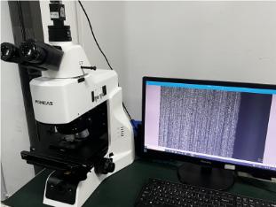
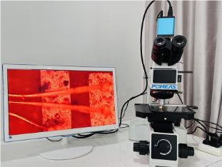
Laser autofocus microscopes leverage their high precision, non-contact measurement capabilities, and rapid imaging to measure surface topography and roughness, three-dimensional structures and dimensions, dynamic changes in living cells, parameters of semiconductor and microelectronic components, as well as material microstructure and composition distribution. They also support multimodal data fusion analysis.

I. Surface Topography and Roughness Measurement

-
Non-contact scanning: Laser autofocus microscopes employ non-contact measurement methods, scanning sample surfaces with laser beams without requiring stylus contact. This eliminates potential damage to samples caused by traditional contact measurements, making it particularly suitable for measuring soft or viscous materials.
-
High-Precision Measurement: With a small laser spot radius (e.g., 0.2μm), it can measure surface roughness in minute areas inaccessible to contact probes, such as narrow regions like fine wires. Simultaneously, its measurement accuracy reaches the sub-micron level, meeting high-precision measurement requirements.
-
Multiple measurement modes: Supports linear roughness measurement (profile method) and surface roughness measurement (surface method). It can measure roughness along a single line or within any rectangular area on the sample surface, providing a more accurate description of the surface condition.
II. Three-Dimensional Structure and Dimension Measurement

-
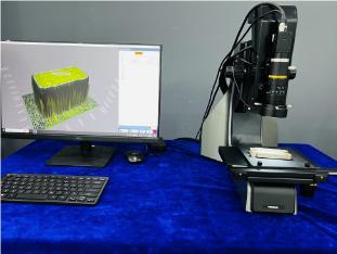
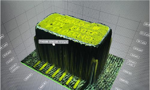
3D Reconstruction: The laser autofocus microscope achieves three-dimensional reconstruction of samples through Z-axis scanning, enabling the acquisition of continuous optical sections across various cross-sections of cells or tissues to form three-dimensional structural images.
-
High-Resolution Imaging: Utilizing advanced confocal technology, it eliminates out-of-focus light interference, enhances imaging resolution and contrast, and provides clearer observation of three-dimensional structures.
-
Dimension Measurement: Enables precise measurement of three-dimensional dimensions such as height, width, and depth, providing critical data support for research in materials science, biomedicine, and related fields.
III. Measurement of Dynamic Changes in Living Cells

-
Long-term dynamic observation of live cells: Laser autofocus microscopy combined with a focus stabilization system enables extended dynamic observation of live cells, ensuring stable focus and preventing blurred imaging caused by sample movement or environmental changes.
-
Quantitative analysis of intracellular biochemical components: Capable of measuring intracellular pH levels and concentrations of various ions (e.g., Ca²⁺, K⁺, Na⁺, Mg²⁺) along with their dynamics, providing a crucial tool for studying intracellular kinetics.
-
Cell Morphometry: Enables precise measurement of cellular morphology parameters including cell area, perimeter, shape factor, and more, delivering quantitative data for cell biology research.
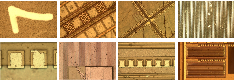
IV. Semiconductor and Microelectronic Component Measurement

-
Wafer Manufacturing Process Inspection: During wafer coating, development and etching, and impurity doping stages, laser autofocus microscopes precisely inspect coating uniformity and thickness, etching depth and width, impurity distribution and concentration, ensuring wafer manufacturing quality.
-
Chip Packaging Process Inspection: During chip packaging, laser autofocus microscopes are critical for inspecting bump height and coplanarity. They enable high-precision measurement of bump height and coplanarity, ensuring reliable bonding between chips and substrates.
-
Microelectronic Component Defect Detection: Rapidly locates minute defects in microelectronic components, such as cracks and scratches, enhancing inspection efficiency and accuracy.
V. Measurement of Microstructure and Composition Distribution in Materials
-
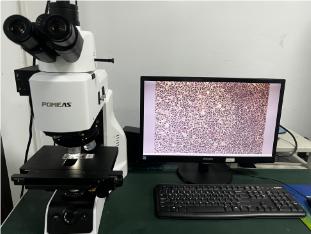
Microstructural Analysis of Materials: Laser autofocus microscopes enable in-depth analysis of material microstructures, including grain size, phase distribution, defect types, and more, providing robust support for optimizing material properties.
-
Composition Distribution Measurement: By integrating techniques such as fluorescent probes or Raman spectroscopy, laser autofocus microscopes visualize the spatial distribution of material compositions, offering a vital tool for materials science research.
VI. Multimodal Data Fusion Analysis

-
Integrated Multi-Imaging Modes: The laser autofocus microscope integrates multiple imaging modes including fluorescence, brightfield, and darkfield. By automatically switching light sources and filters, it achieves multimodal data fusion.
-
Multimodal Data Fusion Analysis: By combining data from different imaging modes, the laser autofocus microscope delivers comprehensive sample information—such as morphological and molecular details, chemical composition, and spatial distribution—providing robust support for analyzing complex samples.
Product recommendation
TECHNICAL SOLUTION
MORE+You may also be interested in the following information
FREE CONSULTING SERVICE
Let’s help you to find the right solution for your project!
 ASK POMEAS
  PRICE INQUIRY
  REQUEST DEMO/TEST
  FREE TRIAL UNIT
  ACCURATE SELECTION
 - APPICATION CASE
- RESOURCE CENTER
- DOWNLOAD CENTER
SOLUTIONS SUPPORT
- ZOOM LENS SELECTION TOOL
- TELECENTRIC LENS SELECTION TOOL
- FA LENS SELECTION TOOL
- ZOOM RATIO TABLE
- CERTIFIED MODEL
SELECTION TOOL
- WHY POMEAS
- FAQ
- PRIVACY POLICY
- TERMS OF USE
- DELIVERY & RETURN POLICY
CUSTOMER CARE
 ADDRESS
Add.:No.68, Chongwei Road, Baizhoubian, East district, Dongguan, China, 523000
CONTACT
 Tel:+ 86-0769-2266 0867
 Fax:+ 86-0769-2266 0867
 E-mail:marketing@pomeas.com

Wechat QR code
Software Copyright :2021SR0176001 抄袭必究, 技术支持:誉新源科技