


How does a super depth-of-field microscope enable high-precision measurements?
Principle of Super Depth of Field Microscope

1. Autofocus scanning and layered imaging
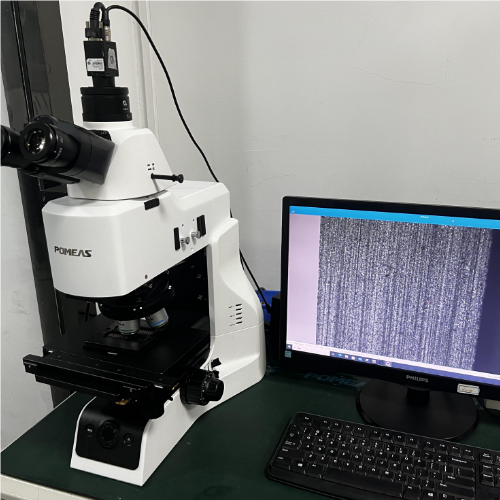
Ultra-depth-of-field microscopes continuously capture images at different focal planes by adjusting the objective lens-sample distance with micrometer precision through stepper motors or piezo-ceramic actuators. For example, the POMEAS PX63L series has a depth of field of up to 32mm at 20x objective, far exceeding the shallow depth of field of conventional microscopes.
2. Multi-focal plane image fusion algorithms
(1) Sharpness synthesis method: the sharpest region is selected for stitching by analyzing the local contrast of each image layer.
(2) Minimum Blur Synthesis Method: based on the evaluation of image blur, the most detailed parts are retained.
(3) High Dynamic Range (HDR) Technology: combines multi-exposure images to solve the problem of overexposure or underexposure of a single image and improve detail reproduction.
Super Depth of Field Microscope Function

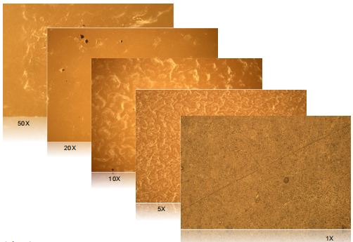
1. The high-resolution imaging system is equipped with a 5-megapixel CMOS sensor that supports continuous zoom from 30X to 5000X to ensure detail capture capability. The optical resolution is up to 500nm, meeting the needs of nanoscale observation.
2. Three-dimensional measurement and topographic analysis:
(1) 2D measurement: label points, lines, circles, angles, arc lengths and other parameters, automatically fit the center line and edge detection;
(2) 3D measurement: calculate height difference between points, volume, surface area and roughness, support profile contour analysis;
(3) Fast 3D modeling: depth synthesis and 3D reconstruction can be completed within 1 second.
3. Intelligent auxiliary functions: AI automatic inspection: performs platform movement, depth synthesis, size determination and defect classification. Multi-position Measurement: Realize multi-point data acquisition through embedded drive device to improve statistical efficiency.
Ultra Deep Field Microscope Applications
- Material analysis: observe the distribution of metal grains, ceramic pores, and plastic fibers with an accuracy of 0.1μm.
- Semiconductor Inspection: ldentify defects on chip surface, measure joint line width, ensure product quality.
- Surface treatment evaluation: analysis of plating thickness, coating uniformity and support for process optimization.
- Cellular imaging: observe cellular 3D structure, neuronal synaptic connections, long working distance design (≥25mm) for easy observation of in vivo samples.
- Histological analysis: synthesize panoramic deep images of thick tissue sections, avoiding the focal distortion of conventional microscopy.
- Pathological diagnosis: assist in identifying tumor tissue boundaries, microbial morphology, and improve diagnostic accuracy.
Ultra-depth-of-field microscope realizes high-precision and multi-dimensional microscopic observation through the combination of optical scanning, algorithm fusion and intelligent measurement technology. Its technical advantages are not only reflected in the hardware performance (e.g. large depth of field, high resolution), but also rely on the software algorithms to deeply mine the image information. It has become an indispensable precision measurement tool in industrial quality control, biomedical research and other fields.
Product recommendation
TECHNICAL SOLUTION
MORE+You may also be interested in the following information
FREE CONSULTING SERVICE
Let’s help you to find the right solution for your project!

 ASK POMEAS
  PRICE INQUIRY
  REQUEST DEMO/TEST
  FREE TRIAL UNIT
  ACCURATE SELECTION
 - APPICATION CASE
- RESOURCE CENTER
- DOWNLOAD CENTER
SOLUTIONS SUPPORT
- ZOOM LENS SELECTION TOOL
- TELECENTRIC LENS SELECTION TOOL
- FA LENS SELECTION TOOL
- ZOOM RATIO TABLE
- CERTIFIED MODEL
SELECTION TOOL
- WHY POMEAS
- FAQ
- PRIVACY POLICY
- TERMS OF USE
- DELIVERY & RETURN POLICY
CUSTOMER CARE
 ADDRESS
Add.:No.68, Chongwei Road, Baizhoubian, East district, Dongguan, China, 523000
CONTACT
 Tel:+ 86-0769-2266 0867
 Fax:+ 86-0769-2266 0867
 E-mail:marketing@pomeas.com

Wechat QR code
Software Copyright :2021SR0176001 抄袭必究, 技术支持:誉新源科技