


The microscope is undoubtedly our most capable partner in the marvelous exploration of the microscopic world. From tiny cells to fine material structures, microscopes allow us to see details that are difficult to detect with the naked eye. POMEAS, as a professional brand in the field of microscope, is committed to bringing you better microscope products to help you explore the microscopic world.

Light microscopy: an introduction to microscopic exploration

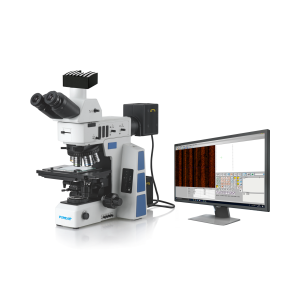
A light microscope is the most common type of microscope and it works based on the refraction of light. Through a series of lenses, the light is focused on the sample and after magnification, we can observe the microstructure of the sample in the eyepiece. This type of microscope is easy to operate and relatively affordable, and is widely used in the fields of education, medical care and basic scientific research. In school biology classes, students observe onion epidermal cells, lacewings, etc. through optical microscopes, opening up the microscopic world of exploration. In hospital laboratories, optical microscopes are used to observe cell morphology in blood and urine samples to help doctors diagnose diseases. According to different application scenarios, optical microscopes can be subdivided into biological microscopes, stereomicroscopes, metallurgical microscopes and so on. Biological microscopes are mainly used to observe biological samples, stereomicroscopes focus on observing the three-dimensional structure of objects, and metallographic microscopes are mainly used to study the microstructure of metal materials.
Electron microscopy: a high-resolution microscopic insider

The advent of the electron microscope has taken our knowledge of the microscopic world to a whole new level. Unlike optical microscopes, it utilizes electron beams instead of light, greatly improving resolution. Scanning Electron Microscope (SEM) is an important member of the electron microscope family, which scans the surface of a sample by emitting an electron beam and collects the reflected back electron signals to generate a high-resolution image of the sample surface. In the field of materials science, SEM can be used to observe the microscopic morphology of metals, ceramics, semiconductors, and other materials, helping researchers to understand the relationship between the properties and structure of materials. Transmission electron microscopy (TEM), on the other hand, is able to penetrate samples and reveal their internal ultrastructure. In biological research, TEM is often used to observe organelles inside cells, the morphology of viruses, etc., providing a key tool for biologists to delve into the mysteries of life.
Fluorescence microscopy: a molecular labeling artifact that illuminates the microscopic
Fluorescence microscopy utilizes the property of fluorescent substances to emit fluorescence under the excitation of light at specific wavelengths to label and observe specific molecules in biological samples. In biological research, fluorescence microscopy plays a pivotal role. Researchers can attach fluorescent substances to specific proteins, nucleic acids and other biomolecules through fluorescent labeling technology, and then observe the distribution and dynamics of these molecules in cells and their interactions under fluorescence microscope. For example, in neuroscience research, scientists use fluorescence microscopy to observe the signaling process between neurons and gain insight into the working mechanism of the brain. In cancer research, by fluorescently labeling specific antigens on the surface of cancer cells, doctors can more accurately detect the presence and spread of cancer cells, providing an important basis for early diagnosis and treatment of cancer.
Atomic force microscopy: a master of microscopic mapping at the nanoscale
An atomic force microscope is a microscope capable of imaging and measuring the surface of matter on the nanometer scale. It measures the change of force between a tiny probe and a sample by means of an extremely weak interaction between the probe and the surface of the sample, thus obtaining microscopic topographical information about the surface of the sample. Atomic force microscopy can be used not only to observe the microscopic undulations on the surface of a material, but also to study the mechanical and electrical properties of the material. In the field of nanotechnology, AFM is an indispensable research tool. For example, in the research and development of nanomaterials, scientists use AFM to accurately measure the size, shape and surface roughness of nanoparticles, optimize the preparation process of nanomaterials, and improve the performance of materials.
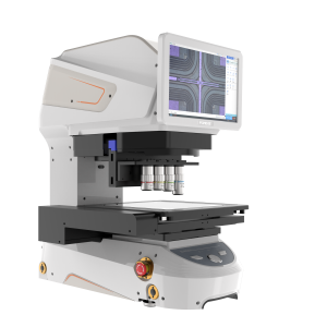
Tool microscopes: precision measurement specialists for industrial manufacturing

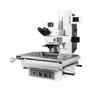
A tool microscope is an optical microscope specialized for precision measurements. It is based on the principle of optical projection, in which the outline or surface details of the object to be measured are magnified and projected onto an observation screen. At the same time, it is equipped with high-precision measuring devices, such as coordinate table, eyepiece micrometer, etc., which can accurately measure the size, shape, position and other parameters of the workpiece. In machinery manufacturing, precision machining and other industries, tool microscope is the key equipment to ensure product quality. For example, in the manufacture of automobile parts, through the tool microscope to measure the dimensional accuracy of engine parts, to ensure the performance and reliability of the engine. In the electronics industry, tool microscope is used to detect the size and shape of tiny electronic components to ensure the quality and stability of electronic products.
Different types of microscopes have their own characteristics, and they play an irreplaceable role in scientific research, medical treatment, industry and other fields. POMEAS provides a wide range of high-quality microscopes to meet the needs of different fields with its professional technology and rich experience. Whether it is a basic optical microscope or a high-end electron microscope, POMEAS is committed to providing users with the clearest and most accurate images of the microscopic world.
Product recommendation
TECHNICAL SOLUTION
MORE+You may also be interested in the following information
FREE CONSULTING SERVICE
Let’s help you to find the right solution for your project!

 ASK POMEAS
  PRICE INQUIRY
  REQUEST DEMO/TEST
  FREE TRIAL UNIT
  ACCURATE SELECTION
 - APPICATION CASE
- RESOURCE CENTER
- DOWNLOAD CENTER
SOLUTIONS SUPPORT
- ZOOM LENS SELECTION TOOL
- TELECENTRIC LENS SELECTION TOOL
- FA LENS SELECTION TOOL
- ZOOM RATIO TABLE
- CERTIFIED MODEL
SELECTION TOOL
- WHY POMEAS
- FAQ
- PRIVACY POLICY
- TERMS OF USE
- DELIVERY & RETURN POLICY
CUSTOMER CARE
 ADDRESS
Add.:No.68, Chongwei Road, Baizhoubian, East district, Dongguan, China, 523000
CONTACT
 Tel:+ 86-0769-2266 0867
 Fax:+ 86-0769-2266 0867
 E-mail:marketing@pomeas.com

Wechat QR code
Software Copyright :2021SR0176001 抄袭必究, 技术支持:誉新源科技