


A Comprehensive Guide to Understanding Autofocus Microscope Classification and Selection
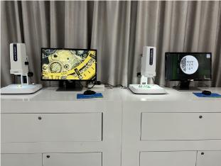
As a core tool for scientific research, industrial inspection, and education, microscopes present users with a dilemma due to their diverse classifications and application scenarios. With the widespread adoption of autofocus technology, selecting the precise model based on specific requirements has become critical. This article begins by examining the classification system of microscopes, analyzes the core implementation logic of autofocus functionality, and provides practical selection guidance for users through application examples of the POMEAS MF Series autofocus video microscopes.

I. Microscope Classification System and Compatibility with Autofocus
Microscopes are primarily classified based on two dimensions: optical principles and application scenarios. Different types exhibit significant variations in their requirements for autofocus technology.
1. Optical Microscope vs. Electron Microscope: The Applicability Boundaries of Autofocus
- Optical Microscope: Utilizing visible light imaging, it is suitable for micrometer-level observation of biological cells, material surfaces, and similar subjects. Autofocus technology significantly enhances operational efficiency in such microscopes. For instance, the POMEAS MF series achieves millisecond-level focusing through real-time image contrast analysis, resolving the time-consuming issues associated with traditional manual focusing.
- Electron Microscope: Relying on electron beam imaging, it achieves nanometer-level resolution, but autofocus requires combining electron beam scanning with feedback algorithms. Such equipment is typically used in high-precision scenarios like semiconductor manufacturing and nanomaterial research, where the autofocus module must be deeply integrated with vacuum systems and high-voltage control.
2. Upright Microscope vs. Inverted Microscope: How Structural Differences Affect Autofocus
- Upright Microscope: The objective lens is positioned above the sample, making it suitable for observing thin specimens such as sections and smears. Autofocus must overcome challenges posed by uneven sample surfaces. For instance, the POMEAS MF series features HDR edge enhancement mode, enabling clear capture of minute defects like scratches on metal surfaces and cracks in ceramics.
- Inverted Microscope: The objective lens is positioned beneath the sample, commonly used for live cell culture and liquid-phase sample observation. Autofocus must adapt to variations in light transmission through containers (such as culture dishes). The MF series employs a four-zone illumination system that allows flexible adjustment of light angles to ensure imaging clarity.
3. Metallurgical Microscope vs Biological Microscope vs Stereo Microscope: Contextual Autofocus Requirements
- Metallographic Microscope: Used for analyzing the microstructure of metallic materials, requiring high-contrast imaging. Auto-focus must be compatible with brightfield/darkfield switching functionality. For example, the MF series supports brightfield (direct reflected light) and darkfield (scattered light) modes, enabling the visualization of features such as grain boundaries and inclusions.
- Biological Microscope: Designed for observing cells and tissues, it must support imaging techniques such as fluorescence and phase contrast. The MF series features a 2-megapixel high-definition camera with 60 frames per second dynamic capture capability, enabling real-time recording of processes like cell division and microbial movement.
- Stereo Microscope: Provides three-dimensional imaging, suitable for electronic component inspection, anatomical observation, and similar applications. Auto-focus requires support for large depth of field and zoom functionality. The MF series motorized ZOOM LENS (0.7x-4.5x) enables rapid switching between observation ranges without changing objectives.
II. Core Technology Implementation of Autofocus Microscopes
The implementation of autofocus relies on the collaboration between hardware sensors and software algorithms, with different technical approaches suited for different scenarios.
1. Image Contrast Method: A Universal Solution
Determining the focal length by analyzing brightness differences in sample images is applicable to most optical microscopes. For example, the MF series employs a CMOS sensor and real-time image processing chip to rapidly identify sample edges and texture features, automatically adjusting to the optimal focus position. This method has low requirements for sample reflectivity but necessitates avoiding interference from highly reflective surfaces.
2. Laser Phase Difference Method: High-Precision Scene Selection
The distance between the objective lens and the sample is calculated using laser reflection signals, making it suitable for complex surfaces such as mirrored surfaces and deep holes. The POMEAS LFS series laser sensors emit laser beams and analyze the reflected signals to calculate distance deviations in real time, driving the adjustment of the focusing module. In three-dimensional morphology inspection of semiconductor packaging solder joints, it achieves a step measurement accuracy of ±0.2μm.
3. Hybrid Focus Technology: Balancing Efficiency and Precision
Combining image contrast with laser phase contrast, the MF series employs image contrast for rapid scanning at low magnifications (2x-20x) and switches to laser phase contrast at high magnifications (50x-100x) to ensure nanoscale detail observation. This mode accommodates the entire process flow, from PCB board flatness inspection to wafer surface etching residue analysis.
III. Selection Guide
1. Magnification and Zoom Range
- Low Magnification (2x-20x): Ideal for rapid scanning of large fields of view, such as PCB board inspection and textile defect screening. The MF Series 0.7x-4.5x motorized ZOOM LENS covers these requirements without changing objectives, enhancing inspection efficiency.
- High Magnification (50x-100x): Used for nanoscale detail observation, such as wafer surface defect analysis. Models supporting long working distance objectives must be selected to prevent lens contact with the sample.
2. Light Source Matching and Imaging Quality
- Multi-Light Source Modes: The MF series offers ring illumination, coaxial illumination, bottom illumination, and other light sources to adapt to different sample characteristics. For instance, ring illumination enhances edge contrast for metal surface inspection, while coaxial illumination reduces reflection interference when observing transparent samples.
- HDR and Edge Enhancement: High Dynamic Range (HDR) technology improves image brightness and contrast, while edge enhancement mode refines minute structures. For instance, the MF series clearly displays cell membrane boundaries during biological cell observation.
3. Interface and Storage Capabilities
- Data Transfer: The MF series supports dual output modes via HDMI, USB 3.0, and LAN, enabling real-time connection to monitors or computers for multi-user collaborative observation and data analysis.
- Storage Methods: An integrated USB port and PC software enable storage of static images and dynamic videos, ideal for quality traceability and failure analysis. For instance, during smartphone case screw hole inspection, the MF series can quickly focus on targets after variable zoom and store defect images for subsequent improvements.
4. Mechanical Structure and Operational Ease
- Integrated Design: The MF series features a compact body that minimizes performance fluctuations caused by external factors, making it suitable for integration into automated production lines or robotic arms.
- Human-Machine Interface: A streamlined touch panel and shortcut key design lower the learning curve. For instance, the MF series supports one-touch storage and light source switching, enabling even non-specialists to operate it quickly.
Product recommendation
TECHNICAL SOLUTION
MORE+You may also be interested in the following information
FREE CONSULTING SERVICE
Let’s help you to find the right solution for your project!

 ASK POMEAS
  PRICE INQUIRY
  REQUEST DEMO/TEST
  FREE TRIAL UNIT
  ACCURATE SELECTION
 - APPICATION CASE
- RESOURCE CENTER
- DOWNLOAD CENTER
SOLUTIONS SUPPORT
- ZOOM LENS SELECTION TOOL
- TELECENTRIC LENS SELECTION TOOL
- FA LENS SELECTION TOOL
- ZOOM RATIO TABLE
- CERTIFIED MODEL
SELECTION TOOL
- WHY POMEAS
- FAQ
- PRIVACY POLICY
- TERMS OF USE
- DELIVERY & RETURN POLICY
CUSTOMER CARE
 ADDRESS
Add.:No.68, Chongwei Road, Baizhoubian, East district, Dongguan, China, 523000
CONTACT
 Tel:+ 86-0769-2266 0867
 Fax:+ 86-0769-2266 0867
 E-mail:marketing@pomeas.com

Wechat QR code
Software Copyright :2021SR0176001 抄袭必究, 技术支持:誉新源科技