Laser autofocus microscopy for life sciences applications - Cell Observation
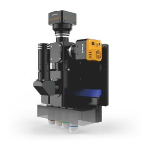
The POMEAS Laser AF Microscope is an advanced microscopic imaging technology that combines laser technology with autofocus capabilities. It provides a powerful tool for fields such as scientific research and industrial inspection by combining laser technology with a high precision autofocus system. It consists of seven modules, including an industrial camera, a coaxial light source, an objective focusing motion module, an objective mounting module, a microscope tube module, a laser sensor module, and an APO objective plate. These modules work together to ensure stable, clear and precise microscopic observation.


Program Background:
In the field of life sciences, cell observation is an important means to study the structure, function and metabolic mechanism of organisms. Traditional microscope observation methods have problems such as long focusing time, low image resolution, and susceptibility to environmental interference, which are difficult to meet the needs of modern biological research for high resolution, high sensitivity and rapid detection. The emergence of laser autofocus microscope system provides a new solution for cell observation.
The system utilizes the high brightness and high resolution characteristics of the laser beam to achieve clear observation of cells. Through the precise control of the laser beam and the autofocus function, the system is able to quickly find and lock the observation target, avoiding the tedious process of frequent manual focus adjustment in the traditional method. At the same time, the non-contact detection method also avoids the contamination and damage that may be introduced by traditional methods, ensuring the integrity and safety of the samples.


Advantages of observing cells:
1. High precision: The laser autofocus microscope system is capable of achieving micron-level detection accuracy, providing higher resolution and finer images for cell observation. This enables researchers to more clearly observe details of the internal structure of cells, organelle distribution and dynamic changes.
2. Non-contact detection: The system utilizes non-contact detection to avoid contamination and damage that may be introduced in traditional detection methods. This is especially important for precious cell samples, which can maintain the original state of the samples and improve the accuracy and reliability of the experiments.
3. Rapid detection: The system has a fast autofocus function, which enables it to complete the task of detecting a large number of samples in a short period of time. This greatly improves experimental efficiency and reduces the workload of researchers.
4. Visualization of detection results: Detection results can be displayed in the form of images or videos, making the detection process more intuitive and easy to understand. Researchers can clearly see the morphology, structure and dynamic changes of the cells, thus analyzing the experimental results more accurately.
5. Multiple Observation Modes and Image Processing Functions: The laser autofocus microscope system supports a variety of observation modes and image processing functions, such as three-dimensional imaging, time series scanning, etc. These functions can meet the needs of different scientific research fields. These functions can meet the needs of different scientific research fields and provide more comprehensive and in-depth analysis means for cell observation.
The POMEAS laser autofocus microscope not only improves the precision and efficiency of observation, but also ensures the integrity and safety of the samples. With the continuous development of the technology, the laser autofocus microscope will play an even more important role in life science research.
Product recommendation
TECHNICAL SOLUTION
MORE+You may also be interested in the following information
FREE CONSULTING SERVICE
Let’s help you to find the right solution for your project!


 ASK POMEAS
  PRICE INQUIRY
  REQUEST DEMO/TEST
  FREE TRIAL UNIT
  ACCURATE SELECTION
  ADDRESS
 Tel:+ 86-0769-2266 0867
 Fax:+ 86-0769-2266 0867
 E-mail:marketing@pomeas.com
